

Optomap Ultra-Widefield
Retinal Imaging
In only a 1/4 second, we can Image
82% of the inside of your eye.
What is OPTOMAP Retinal Imaging?
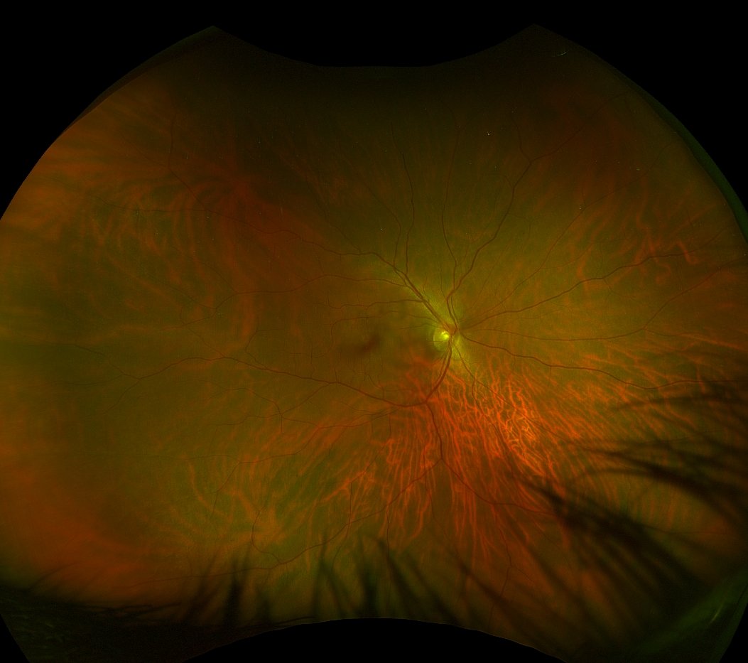
Optomap ultra-widefield digital imaging is an exciting technology that allows you and your optometrists to see a 200-degree picture or image of the inside of your eye or about 82% of the retina. Traditional examination techniques only allow your eye doctor to examine 10-15% of your retina without dilating your eyes.
This ultra-widefield digital image provides a ‘map’ that helps to guide your optometrist during the eye exam towards areas of concern. This imaging technology is also invaluable in patient education. It allows our optometrists to show you areas of concern that might need monitoring or treatment. It also allows our optometrists to detect even the earliest signs of diseases that appear on your retina.

OPTOMAP Images are a great educational tool that we use to help you to better understand what we're looking at, and looking for.
Why OPTOMAP Is More Than Just a Picture
The California af was the first OPTOMAP to incorporate composite colour and digital autofluorescence imaging. You can see an example below of the difference in the digital images. The autofluorescence digital image provides another tool to determine the cause of underlying vision complaints. It allows our optometrists to uncover problems that would previously go undiagnosed or unexplained.

Composite Colour Imaging
Composite colour digital imaging uses multiple lasers to image the back of the eye. These lasers penetrate the layers of the retina to different depths, allowing your optometrist to visualize and locate the layers of the retina more easily. This aids in the detection, diagnosing and monitoring of various eye conditions. The Green (532nm) laser visualizes the sensory retina to the RPE layer. The Red (635nm) laser looks deeper between the RPE to the choroid. When combined, they produce the digital image you see above, which can be used to help your optometrist explain your eye health.

Autofluorescence Imaging
The auto-fluorescence (AF) mode on the Optos California model helps our optometrists monitor retinal conditions by showing abnormalities or metabolic changes within the retina that may not be visible during standard digital retinal photography or in a dilated retinal exam. This test is valuable in patients with conditions such as diabetic retinopathy, age-related macular degeneration or hydroxychloroquine toxicity because it can help determine if the affected area is in an active or stable phase. In addition, this non-invasive test can regularly be performed on patients of all ages.
How does autofluorescence work?
OPTOMAP af (autofluorescence) is a non-invasive, in-vivo imaging modality used to provide information on the health and function of the retinal pigmented epithelium (RPE). Over time, the retinal photoreceptors naturally age and produce metabolic waste known as lipofuscin. Lipofuscin is the fatty substance found in the retinal pigmented epithelium. Excessive amounts can be caused by the aging retina, certain retinal diseases and the progression of diseases. It has been thought that excessive levels of lipofuscin could affect essential RPE functions that contribute to the progression of age-related macular degeneration (AMD). These findings have also been shown to have prognostic value and help predict which eyes are at a greater risk of advanced disease progression.
Typical, autofluorescence imaging is clinically useful in age-related macular degeneration, central serous retinopathy, choroidal tumours and birthmarks known as nevi, inflammatory diseases, inherited diseases, optic nerve head drusen, retinal toxicity and retinal detachments.
We Include Optomap Imaging as Part of Every Comprehensive Eye Exam.
Does OPTOMAP Replace a Dilation?
Although OPTOMAP imaging provides our optometrists with an ultra-widefield view of your retina, it does not replace the need for a regular dilated retinal exam. Patients with conditions such as diabetes, hypertension, glaucoma, and age-related macular degeneration still require this test on an annual basis, as it's still considered the standard of care. A dilated eye exam provides more detail to your optometrist by understanding elevations and depressions, clarity of the eye, and a more dynamic view of the retina.
Optomap ultra-widefield images are simply a tool our optometrists use to help uncover and monitor eye diseases during an eye exam. They can also improve a dilated eye exam by giving your doctor a map of where to focus their examination.
OPTOMAP FAQ’s
-
The ultra-widefield optomap may help your eye doctor detect problems more quickly and easily. In addition, unlike traditional retinal exams, the optomap images can be saved for future comparisons. They also allow you to see what your optometrist sees, making your eye exam more interactive and educational.
-
We recommended that you have an OPTOMAP image taken as part of every routine eye exam. We may perform additional images if we’re monitoring specific eye conditions, or if you develop visual concerns between visits.
-
OPTOMAP images are created by non-invasive, low-intensity scanning lasers. No adverse health effects have been reported in over 150 million patient visits. However, OPTOMAP does create a flash, which can trigger a photosensitive seizure in people prone to this. So if you've ever had problems with flashing or flicking lights, we may elect not to perform this test.
-
Yes. In fact, many vision problems begin in early childhood, so it's important for children to receive quality routine eye care. At Helio Optometry, we start using OPTOMAP imaging as soon as children can perform the test, usually between ages 4-5. Kids find the images fascinating, and it helps them to understand how their eyes work.